Steps for the Work flow
STEP 1: UniProt – Protein Identity & Function (Starting Point)
UniProt is the central hub that links all other protein resources. You can get the protein name, gene, organism, function and biological role, domains and active sites, disease relevance, and cross-references to PDB, AlphaFold, and Human Protein Atlas (HPA).
- Go to https://www.uniprot.org
-
Search by:
- Gene name (e.g., TP53)
- Protein name (e.g., Insulin)
- Accession number (e.g., P01308)
Example (TP53 – P04637):
- Function: Tumor suppressor, transcription factor
- Disease: Cancer-associated mutations
- Links available to PDB, AlphaFold, and Human Protein Atlas
STEP 2: PDB – Experimental 3D Structure
PDB provides experimentally determined structures using X-ray crystallography, Cryo-EM, and NMR spectroscopy. You can obtain information on protein folding and domains, binding pockets and active sites, ligand interactions, and the structural basis of function.
How to Navigate
- From the UniProt entry → scroll to Cross-references
- Click PDB
- Open a structure in RCSB PDB
Example (Insulin): PDB shows insulin’s disulfide bonds, explaining stability and biological activity. If a structure exists in PDB, it is the gold standard.
STEP 3: AlphaFold – Predicted Protein Structure
Why AlphaFold Is Needed
Many proteins do not have experimental structures. AlphaFold fills this gap with high-accuracy predictions. You can obtain information about overall protein fold, confidence regions vs flexible regions, and structural hypotheses for function.
How to Use AlphaFold
- From UniProt → click AlphaFold DB link
- View interactive 3D model
-
Examine pLDDT confidence scores:
- High pLDDT (>90) = very reliable prediction
Example (BRCA1):
- No full-length PDB structure available
- AlphaFold predicts full protein architecture
- Low-confidence regions indicate intrinsic disorder
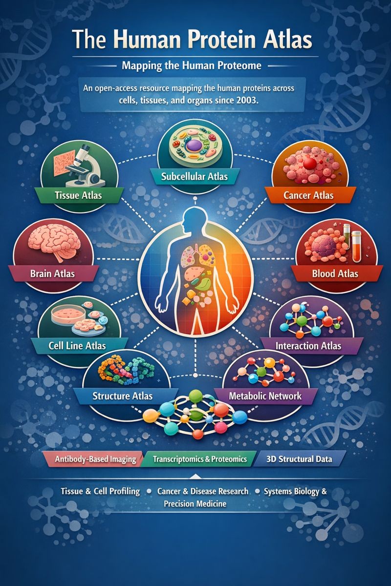
STEP 4: Human Protein Atlas (HPA) – Expression & Localization
Why Use HPA?
Structure alone is not enough. HPA answers: Where is the protein expressed? In which tissues and cell types? Is it linked to cancer or disease prognosis? You can get information about: Tissue-specific expression; Subcellular localization (nucleus, cytoplasm, membrane); Protein presence in normal vs cancer tissues and Prognostic significance. So overall structure explains how → HPA explains where and when.
How to Navigate
- Go to https://www.proteinatlas.org
- Search by gene name (e.g., TP53)
-
Explore:
- Tissue Atlas
- Cell Type Atlas
- Subcellular Atlas
- Cancer Atlas
Example (TP53):
- High nuclear expression
- Elevated in multiple cancers
- Strong clinical relevance
STEP 5: Integrating All Resources (Systems Understanding)
This integration creates a unified model that connects a protein’s sequence, structure, and tissue expression to decode disease at a systems level. Together, these resources accelerate biomedical discovery, transforming genetic clues into precise insights for diagnosis and therapy.
Example: Insulin (INS)
| Resource | Insight Gained |
|---|---|
| UniProt | Function in glucose metabolism |
| PDB | Disulfide-bonded structure |
| AlphaFold | Complete precursor structure |
| HPA | High expression in pancreatic β-cells |
Conclusion: Structure supports hormone function, expression confirms endocrine specificity.
STEP 6: Using BLAST Across the Workflow
Purpose
BLAST allows the study of evolution and conservation.
Workflow Integration
- BLAST sequence from UniProt
- Identify homologs
- Compare AlphaFold structures
- Compare tissue expression across species (HPA / literature)
Example: BLAST insulin → compare human vs fish insulin → conserved function despite sequence variation.
STEP 7: Workflow Checklist
- ✔ Identify protein → UniProt
- ✔ Analyze function & domains → UniProt
- ✔ Examine structure → PDB
- ✔ Predict missing structure → AlphaFold
- ✔ Verify tissue & disease expression → Human Protein Atlas