
Mitochondria
Mitochondria originated approximately 1.5–2.0 billion years ago when an ancestral eukaryotic cell engulfed an aerobic alphaproteobacterium, which evolved into the mitochondrion. The mitochondrion supported high-energy metabolism that enabled complex eukaryotic cells and later multicellular organisms.
This evolutionary origin explains why mitochondria have their own DNA (mtDNA), separate from nuclear DNA, and why they retain bacterial-like features such as circular DNA and their own ribosomes for protein synthesis.
Every cell contains hundreds to thousands of mitochondria (except red blood cells), with oocytes containing over 100,000. Oocytes have very high mtDNA copy numbers. However, during oogenesis and early development, only a relatively small effective set of mtDNA molecules seeds the next generation (the mitochondrial genetic bottleneck), which can shift heteroplasmy levels between mother and child.
Mitochondria form dynamic, interconnected networks that constantly undergo fission (splitting) and fusion (combining)—processes essential for health and function. Fission allows damaged mitochondria to be segregated and removed (mitophagy), while fusion enables mixing of contents to maintain functional integrity. Disruptions in these processes cause specific mitochondrial diseases.
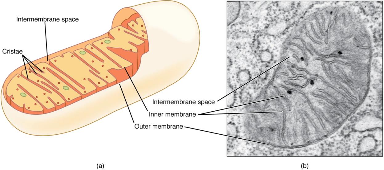
Structure of Mitochondria
Outer Membrane
The outer membrane is relatively permeable and interacts with the endoplasmic reticulum (ER) to regulate calcium. Its permeability comes from porin proteins that allow small molecules to pass freely. Its close association with the ER creates specialized contact sites where calcium is transferred between organelles, crucial for cellular signaling.
Inner Membrane
The inner membrane is highly impermeable to most ions, which is essential for maintaining the proton gradient. This property is supported by its lipid composition, including cardiolipin, and specialized transporters. Cardiolipin makes the membrane virtually impenetrable to ions—essential for maintaining the proton gradient.
Cristae increase the inner membrane surface area several-fold, enabling dense packing of OXPHOS complexes.
Intermembrane Space
The region between outer and inner membranes accumulates protons pumped during electron transport, creating an electrochemical gradient. It also contains cytochrome c, which is released during apoptosis initiation.
Matrix
The central space contains mitochondrial DNA, ribosomes, and metabolic enzymes. The matrix is where the Krebs cycle operates, where fatty acid oxidation occurs, and where mtDNA is transcribed and translated— making it the metabolic headquarters of the organelle.
Image Source: "OpenStax Anatomy and Physiology (CC BY 4.0). Access for free at Check here
The Oxidative Phosphorylation (OXPHOS) System
The inner membrane houses five protein complexes (I–V) that work together to generate ATP through oxidative phosphorylation. This system functions like a cellular power plant.
- Complexes I–IV: Transfer electrons from NADH and FADH₂ to oxygen while pumping protons across the membrane. Electrons enter through Complex I (from NADH) or Complex II (from FADH₂), then flow to coenzyme Q10, Complex III, and cytochrome c. Complex II transfers electrons to CoQ but does not pump protons. Finally, at Complex IV, oxygen accepts electrons to form water. Each transfer releases energy used to pump protons.
- This process creates an electrochemical gradient (like water behind a dam). The gradient has two components: a pH difference (alkaline in the matrix) and a voltage difference (negative inside), together called the proton motive force.
- Complex V (ATP synthase) allows protons to flow back across the membrane, capturing that energy to synthesize ATP from ADP and phosphate. Protons flowing through the rotor subunit cause it to spin like a turbine, mechanically driving ATP synthesis—a remarkable nanoscale biological machine.
Other Functions of Mitochondria
Mitochondria also regulate:
- Calcium Homeostasis: Mitochondria take up calcium through the mitochondrial calcium uniporter, buffering cytoplasmic calcium levels and modulating signaling pathways. This function is particularly important in neurons and cardiomyocytes.
- Apoptosis: When mitochondria receive death signals, they release cytochrome c and other factors that activate caspases— executing the cell’s programmed suicide mechanism. This positions mitochondria as central decision-makers in cell survival.
- Reactive Oxygen Species (ROS) Management: As byproducts of electron transport, superoxide is produced primarily at Complexes I and III. Mitochondria contain intrinsic antioxidant systems such as manganese superoxide dismutase and glutathione peroxidase to prevent oxidative damage.
- Multiple Metabolic Pathways: The mitochondrial matrix participates in numerous metabolic processes. The urea cycle is compartmentalized, with its initial steps (including carbamoyl phosphate synthetase I and ornithine transcarbamylase) occurring in the mitochondrial matrix, while subsequent reactions take place in the cytosol. This dual localization highlights the integrated nature of mitochondrial–cytosolic metabolic coordination.
In addition, mitochondria contribute to heme synthesis, steroidogenesis (shared with the smooth endoplasmic reticulum), fatty acid oxidation, and iron–sulfur cluster assembly.
Mitochondrial DNA (mtDNA)
Mitochondria possess a unique feature: they contain their own DNA known as mitochondrial DNA (mtDNA), which is separate from the nuclear DNA present in the cell nucleus.
The Mitochondrial Proteome
Mitochondria contain approximately ~1,150 proteins, yet only 13 proteins are encoded by mtDNA. The remaining proteins are nuclear-encoded and imported into the organelle through specialized translocase systems known as the TOM (Translocase of the Outer Membrane) and TIM (Translocase of the Inner Membrane) complexes.
MitoCarta (e.g., MitoCarta 2.0 and later updates such as MitoCarta 3.0) serves as a high-quality compendium of mitochondrial proteins. It is compiled using integrated experimental and computational evidence including:
- Proteomics data
- Microscopy-based localization studies
- Targeting sequence prediction
- Literature curation
Rather than relying on a single validation method, MitoCarta aggregates multiple lines of evidence to support mitochondrial localization. This makes it an essential reference for gene prioritization and variant interpretation in mitochondrial disease genomics.